Sunday, February 19, 2012

The Cost of Compensation

So, we've discussed a few of the ways that the body knows the blood pressure and blood flow has changed, and also how it goes about making those changes.

It the arteries, the baroreceptors that exist in the aorta and carotid arteries sense changes in blood pressure (which the communicate to the brain).  If a change needs to occur, it can be a nervous change, an endocrine (or hormonal) change, or a structural change (in order of speed).  All matters of change could occur, we are only listing the types.

In regards to blood flow, the system is less developed.  Direct changes occur in tissues, known as metaboreflexes.  Metaboreflexes occur in response to changes in oxygen, nutrient and waste levels in the tissue and cause vasodilation or vasoconstriction.  When vasodilation or vasoconstriction has a profound effect at the heart, indirect changes as a result of changes in blood flow occur, piggy-backing on the baroreceptors of the blood pressure detection system to cause big changes.

Something we haven't discussed is how changes in the veins are measured.  Well, there are technically baroreceptors in the major veins as well.  However, they are much more forgiving of change and are not termed as baroreceptors to avoid any confusion with the more important baroreceptors of the arteries.


So, that leads us to our final consideration (for now) when thinking about these reflexes of change in blood pressure or blood flow.  What is the cost?  Well, actually, it's good that you ask!  There is always a cost to compensation in the CV System.  


For example, when changes in the nervous system occur, what are the costs?  Well, if the sympathetic nervous system is stimulated to increase heart rate and contractility to provide more oxygen (via blood) to tissues, what has to work harder?  Right!  Both the heart and the lungs have to work harder.  Increasing the work of either tissue is going to increase the amount of waste that tissue produces.  This, in the long term, can result in damage to the tissues, which can later lead to apoptosis (or cell death).

How about the negative consequences of renin activation in the endocrine system?  Well, when renin increases, it not only leads to salt retention in the kidneys, but it also leads to the production of the hormone angiotensin II in the kidneys.  Guess what, chronic increases in angiotensin II levels can lead to deterioration of the heart via the hormone triggering remodeling.  


Finally, how about hypertrophy?  First and foremost, hypertrophy is known to shorten the lifespan in humans.  Why?  Well, hypertrophy, overtime, is going to cause less flexibility in the heart wall.  This means that preload and afterload are going to have to increase to overcome the increased pressure required for the heart to either fill or contract (for ejection of blood).

As you can see, in the short term, these adaptations are essential to keep the animal alive.  However, in chronic situation, all of these adaptations come with consequences.  These consequences (if sustained) will eventually lead to heart disease if not controlled.

- JD

What if there's a problem with Flow?

So, we just got done talking about the baroreceptors.  The baroreceptors monitor the stretch that exists in the aorta and carotid arteries, and then it delivers a message about the changes to the brain via nerves.  Basically, then, we have described changes about the amount of blood present, or (more specifically) changes about the blood pressure.

Well, how about changes in blood flow (which we will just talk about the biggie, cardiac output)?

Before we get too far, just know, the system of detection concerning blood flow is much less developed than the system that detects changes in blood pressure.

Generally, blood flow changes are more so detected in individual tissues or organs.  As the body is constantly changing to ensure that the most important organs are receiving blood, this is factor one.  Next thing to think about, the body is vast!  There are plenty of places that blood flow could be slowed.  So, then, how does the body react to changes in blood flow?

Unfortunately, there are no receptors that are going to detect an increase or decrease in blood flow (per se).  Instead, changes in blood flow are detected (and then adjusted) by an organ itself.  So, for example, perhaps with have a thrombus (or blood clot) in the right kidney.  Oh no!  With there being a blood clot in the right kidney, that means that the right kidney is going to be receiving less than adequate flow of blood due to blockage.  When less than adequate blood is delivered to an organ, the organ tissues become ischemic (a term that describes a lack of adequate blood to a tissue).  In response to ischemia, a series of events will occur, but most importantly vasodilation will occur.  We discussed vasodilation, do you remember what that means?  Right!  The blood vessels will dilate (or expand) to cause more blood to flow to an area.  In our case, the blood vessels will be vasodilating to cause more blood flow to the right kidney.  

In the grand scheme of things, vasodilation to just that one right kidney is going to have that much of an impact on blood flow to the heart or brain.  But, how about in a case where thrombi have clogged the right kidney, the liver, and the large intestine.  Holy cow!  The vasodilation that results in the right kidney, liver and large intestine (with the liver and large intestine being much larger organs that the right kidney) is likely to make a much more profound impact at the heart and brain.  Why?  Well, if more blood is flowing to the right kidney, liver and large intestine, where did that blood come from?  It had to have taken from the blood that is flowing to the heart and brain.  So, when less blood than expected returns to the heart and brain, those organs are going to notice!

How do you suppose they know that there is less blood returning?  That's right!  If there is less blood returning to the heart, that means there will be less stretch in the baroreceptors (indicating that blood pressure has fallen).    

So, there are two methods by which a change in blood flow is measured.  The first is a direct measurement of change in blood flow.  This occurs locally in organ tissues.  Direct detection occurs via detection of oxygen, nutrient and waste levels, all managed via blood flow.  Changes in these levels can cause a tissue to vasoconstrict or vasodilate in response (known as metaboreflexes).

The next was in an indirect measurement.  If metaboreflexes have occured (or, if tissues have vasodilated or vasoconstricted due to changes in oxygen, nutrient or waste levels), the subsequent change in blood pressure is going to be detected by baroreceptors, which communicate to the brain to cause nervous, endocrine or structural changes in response.

So, what's the big picture?  In regards to the detection of changes in blood flow, the system is not as advanced as the system of baroreceptors that detect changes in arterial blood pressure.  However, though weaker, the system that detects changes in blood flow can get big effects once metaboreflexes result in changes of blood pressure that stimulate the baroreceptors, recruiting the more powerful system for assistance.

Cheers! - JD

How the CV System "Knows" there's a Problem

But, before we go there, what were those top Three priorities of the CV System again?
How about the top three equations when discussing the CV System?
Do you remember the equation that allows you to assess wall stress?
How about hypertrophy?  What can you tell me about that?
Finally, can you give me a brief summary in regards to the PV Loop of the LV?

To allow you a moment to marinate on these questions, here is a random screencap of one of my favorite movies:


That's right, Toothless!  And, yes, I'm too old for this cartoon.  But, I just cannot help but feel a little love for this dragon!  Such a nice story for children.  But, I digress....

The Top Three Priorities of the CV System: 


  • Maintain Arterial Blood Pressure
  • Maintain Cardiac Output
  • Maintain Venous Pressure

Great!  The Top Three Equations in the CV System:
  • ArtBP ≈ TPR * CO
  • CO = HR * SV
  • SV ≈ Preload, Afterload, and Contractility
Excellent, again!  The equation for wall stress:

Wall Stress = (Intracavitary Pressure * Radius)
                      Wall Thickness

Outstanding!  How about Hypertrophy?

Hypertrophy describes an increase in cardiac mass.  Hypertrophy can occur in one of two ways, either Concentric Hypertrophy or Eccentric Hypertrophy.  Eccentric Hypertrophy describes an increase in cardiac mass that has an increase in volume but does not have an increase in wall thickness.  Concentric Hypertrophy describes an increase in cardiac mass that has an increase in wall thickness, and either has no change or very little change in cardiac volume.  Hypertrophy, in particular Concentric Hypertrophy, is the easiest way by which the heart can return wall stress to a normal value once increased.

Fantastic job!!  Finally, how about a quick summary of the PV Loop of the LV?

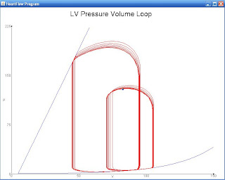
The PV loop is the depiction of a single heart beat in the body.  Along the x-axis, volume is depicted.  Along the y-axis, pressure is depicted.  The line that originates from the (0,0) point on the graph and connects to the Upper Left corner of the PV Loop is a depiction of contractility.  Beginning with the Upper Left corner, this is the point at which the Aortic Valve closes.  The Bottom Left corner is the point at which the mitral valve opens. The vertical line that connects the Upper Left and Bottom Left corner depict the period of isovolumetric relaxtion in the heart (a point of relaxation that has no change in volume).  The Bottom Right corner is the point at which the mitral valve closes.  The Upper Right corner is the point at which the aortic valve opens.  The vertical line that connects the Bottom Right corner to the Upper Right corner depicts the period of isovolumetric contraction in the heart (a point of contraction that has no change in volume).  Starting that the Upper Left corner and ending at the Bottom Right corner, diastole has occurred.  Starting at the Bottom Right corner and ending at the Upper Left corner, systole has occurred.  The width of the PV Loop corresponds to stroke volume and is easily influenced by change in preload, afterload and contractility.  Something to remember is that the PV Loop is not a "snapshot" but rather a line that is traveled to depict a single heartbeat.  The pressure is constantly changing along the PV Loop, while the volume has two points (isovolumetric relaxation and isovolumetric contraction) at which the volume is unchanging.  Also, the PV Loop is incapable of accounting for wall thickness changes, as it only depicts one heartbeat and changes in wall thickness occur over a long period of time. 


And there you have it!  Great job answering those review questions!  They will be important to keep in mind as we proceed through our studies of the CV System!

So, where are we today?  Well, using all of the knowledge from the review questions, what two issues to you feel dominate the CV System/  You've got it, blood pressure and blood flow!  So, how do you suppose the body keeps such tight control over either of these two issues?  Well, as you're about to see, there are many intricate ways in which the body (read: brain) ensures that changes in blood pressure or blood flow are addressed as quickly and diligently as possible.  You see, it must ensure that Priority #1 is maintained, otherwise it will be incapable of controlling any changes that when ensure the body stays alive

In regards to ArtBP, a series of receptors (known as baroreceptors) exist in the body.  Well, what are baroreceptors?  Baroreceptors can be best thought of a cells that detect stretch in the arteries.  How stretched (or distended - dictated by how full of blood the vessel is) the artery is results in a nervous signal being sent to the brain to communicate that stretch.  Let me see if I can give you an example...  Pretend it's Thanksgiving.  What do most people do at Thanksgiving?  They eat, and they eat a lot!  So, as your stomach fills with food, it becomes distended.  Now, what happens when you overeat?  You feel terrible!  With good cause, too.  If you we unaware that your stomach was painfully full (or distended) with food, your stomach might burst (and, for the record, it really would take a whole lot of food to cause this to happen).  Well, there are stretch receptors on your stomach that tell the brain that the stomach, "Hey!  The stomach is too full, stop eating."  What about the opposite?  What happens when there is no stretch in the stomach?  That's right!  Those stretch receptors say, "Hey Brain!  There is no stretch in the stomach, you need to find food!"

Now, the stomach receptors aren't baroreceptors (the baroreceptors exclusively measure the stretch in blood vessels), but the idea is the same.  When the arteries are overstretched because they are too full of blood, a message is sent to the brain that says, "Hey!  Brain, better make some adjustments in the heart (via CO, SV, Preload, Afterload, Contractility) to decrease the stretch in the baroreceptors.  Just the opposite is true too.  When there is not enough stretch in the baroreceptors, the baroreceptors send a message to the brain that says, "Hey!  The arteries are not full enough!  Better make some changes!"  

Now, baroreceptors are not in all arteries.  Actually, the baroreceptors are located in specific arteries.  There are the aortic baroreceptors, in the aorta, and the carotid baroreceptors in the carotid artery

For simplicity, I have edited this photo to highlight only the location of the aorta and carotid arteries.  See how close they are to the heart?  This is much of the reason that these arteries were chosen to possess the baroreceptors.  Remember, Priority #1 is designed to ensure that ArtBP gets blood to the brain and heart (very important organs).

So, how can the brain respond, then?  We've already mentioned causing a change in CO, SV, Preload, Afterload, and Contractility.  But, how?  How does the brain cause those to change?  Well, basically, there are three types of changes that can occur:
  • Autonomic nerve changes
  • Endocrine (or hormonal) changes
  • Structural changes

Starting with the autonomic nerve changes:  It's important to establish that there are two different types of autonomic nerves in the body.  There are the sympathetic nerves, while the others are the parasympathetic nerves.  What's the difference?  Well, the sympathetic nerves control your "Fight or Flight" response.  Well, what does that mean?!  If you encounter a monster, what would you hope to have happen?  Well, you can either fight the monster or you can run fast (of flee) from the monster.  In order for you fight or flight to be successful, you will need a series of things to change in your body to help that out.  Can you think of some things that would help you fight or fly better?  Well, you probably need to breathe faster (to get more oxygen), you will need you heart to pump faster, so that the blood is getting oxygen and nutrients, to the tissues quicker.  You might need your pupils to dilate (or expand) so that you can see more.  And, you definitely don't want to have to use the restroom.  So, you'd like for your intestinal motility to decrease and for you bladder to stay relaxed (rather than contracted, which is what occurs when you urinate).  All of those things, those are the things the the sympathetic nervous system (SNS) causes to happen (or, those are the things the SNS controls). 

The other system is the parasympathetic nervous system (PNS).  The PNS does just the opposite of everything I have listed above.  So, the PNS would reverse those changes once you are done fighting or flying from the monster.  Does that make sense?  

Back to the heart, as you can imagine, when the baroreceptors sense that there is too much stretch in the arteries, the PNS is activated to cause a decrease in CO, SV, Preload, Afterload, or Contractility.  But, wait!  I thought Priority #2 was to maintain Cardiac Output, not decrease it!!!  Well, it is.  You see, if the baroreceptors are too stretched this means that there has already been an unwanted increase in CO or ArtBP.  Yeah?

Now, autonomic nerve changes are by far the quickest response to a change in baroreceptor input.  Followed by those changes, you have endocrine (or homonal) changes.  These, certainly, are slower than any nerve change.  But, they aren't too bad.  The hormone epinephrine is one that will increase heart rate.  The hormone renin is going to affect how the kidneys function.  Again, the kidneys??  What do they have to do with anything?  Well, if the kidneys are triggered by the hormone renin, there are going to excrete (or get rid of) less salt.  For now, just know that water follows salt.  So, if salt is retained by the body, so is water.  This water can then be used to increase blood volume.  There we are, back to the heart!  So, for example, if the baroreceptors sense the the arteries are too empty, the brain may cause a release of renin, which will cause the kidneys to conserve salt (and therefore conserve water), which will end in the increase of blood volume (or, pretty much, result in there being more blood in the arteries).  

Finally, we have structural changes.  Well, we've already talked about this!  You know that hypertrophy causes an increase in cardiac mass.  You know that it can happen in two different way, but you also know that it takes a long time.  Therefore, structural changes are the slowest changes that will occur in response to a change in baroreceptor stretch (and, while I'm here, structural changes in the heart are going to be initiated by the brain through both nervous change and endocrine change).  

So, why three different responses to a change in the baroreceptor, isn't that inefficient?  Well, actually, it is very important for the body to have this three-tier system in response to baroreceptor changes.  You see, each time you sit, stand, exercise, get stressed, there is a change in all of the factors of the CV System (meaning CO, SV, Preload, Afterload, Contractility).  These changes are normally controlled by the nervous change.  In more chronic situations, the endocrine changes happen initially, followed by structural changes.  If it weren't for this three-tier response to change, each time you sat or stood, a structural change could be initiated and cause hypertrophy!  Well, that wouldn't be good at all!

As you can see, though complex, the CV System is intricate for a very important reason:  Not only does it keep you alive, but it also prevents unwanted or unneeded changes from occurring)! 

Cheers! - JD

Pressure-Volume Loops

Pressure-Volume Loops (PV Loops) are a very useful tool during hypothetical analysis of a chamber of the heart.  This refers to any chamber of the heart (RA, RV, LA, or LV).  This will be a picture-heavy post, but let's get a better understand of just what the PV Loops is designed to tell us!

Let's just start with a quick look at what a PV Loop looks like:


There it is in its glory,  Not much to look at, eh?  Don't worry!  With the use of my laptop, let's get a few things labelled so that the PV Loop is a lot less mystery and a lot more awesomely useful.

 Let's start with the axes:


Just for clarity, the x-axis depicts volume, while the y-axis depicts pressure.


This line shown in the PV Loop is a depiction of contractility.  Shortly, you will get a chance to see how a change in contractility (via that line) alters the shape of the PV Loop


The width of the PV Loop corresponds to stroke volume.  Just like contractility, you will shortly see how easily the stroke volume can be altered by the shape of the PV Loop.

Now, for big concepts:  A PV Loop depicts the events of a single, hypothetical heartbeat.  What is also true is that the PV Loop is not a 'snapshot' of the heart.  To read the PV Loop, you must travel along the PV Loop.  Does that make sense?  What I mean is, you pick a point to start (let's say the Upper Left corner, and then follow the loop's line.  Something that is true is that throughout that heartbeat, the pressure is always changing.  As you follow the line of the PV Loop, you never just stay at the same value along the y-axis (the axis that depicts pressure).  You may return or have similar pressure value eventually, but you are just stuck at one particular value of pressure.  How does this apply to the x-axis (the axis that depicts volume)?  Excellent!  There are two points in time in which the volume in the heart is unchanging (depicted by the two vertical lines, one on the left and another on the right of the PV Loop).  There are two points in time (within the heart) that the volume is unchanging, one (the left) value is isovolumetric relaxation and the other (the right value) is isovolumetric contraction.  We will come back to both, shortly.


Here are some of the major events within the left ventricle.  Remember, that the PV Loop can depict activity in any of the four chambers of the heart.  In our example, we are talking specifically about the PV Loop in the Left Ventricle.  Let's start in the Upper Left corner of the loop.  At this moment, I have denoted that the aortic valve closes.  The aortic valve is the valve that exists in the aorta, and can allow or prevent communication between the LV and the body.  As a result of the aortic valve closing, the chamber exists in a state in which no valves are open.  I will follow with a labelled picture, but the left vertical line of the PV Loop depicts, then, a time of isovolumetric relaxation.  Picking apart the word, isovolumetric relaxation refers to a period of relaxation in the heart in which there is no change of volume occurring (a result of the fact that no heart valves are open).

At the Bottom Left corner of the PV Loop, the mitral valve opens.  Remember, the mitral valve is the valves that separates the left atrium from the left ventricle.  What do you suppose happens when the mitral valve opens and allows communication between the LA and the LV?  Of course!  When the mitral valve opens blood is allowed into the left ventricle and diastolic filling of the chamber begins!  Great job!

At the Bottom Right corner of the PV Loop, the mitral valve closes.  Here we are again, with none of the heart valves that communicate with the LV being open.  Again, no change in volume occurs, making this isovolumetric.  This time, however, rather than relaxation occurring, contraction is occurring.  What's the difference, or what makes one relaxation and the other contraction?  I'm glad you asked!  So, let's start with isovolumetric contraction (the right vertical line).  What has occurred in the heart (due to diastolic filling)?  It's filled with blood!  Something we have yet to discuss is that there is electrical activity going on within the heart (preparing the heart to contract for a heartbeat).  So, as the cardiac myocytes begin to contract (or, shrink in size), that pressure, combined with the pressure created by the stretching of cardiac myocytes due to being filled with blood, continually increases.  In contrast, isovolumetric relaxation (the left vertical line) is occurring after contraction (or ejection of blood from the heart) has occurred.  As you can imagine, that is a huge relief or a relaxing event for the heart.  It's allowed to return to its normal, original size.   I will be providing a labelled diagram of this in just a sec, but I think the next paragraph will help here.

Finally, at the Upper Right corner of the PV Loop, the aortic valve opens.  Now, what happens in the heart?  Right!  When the aortic valve opens (alongside that contraction of the left ventricle) blood is ejected from the heart to the aorta (to be carried to the body).  Starting the Bottom Right corner and ending with the Upper Left corner, we have described contraction (or the period of systole in the heart).

Now, for some labelled diagrams:


And, to just be even more helpful (I know my artwork is terrible), here's a professional depiction of the PV Loop, with all of the bits and pieces labelled (I just wanted to go slow, initially).  If you are confused (as I feel that I may not have done as well as I could have, please leave a comment):


So, know, let's look at how fairly isolated changes in preload, afterload and contractility can affect the appearance of the PV Loop.

Here's a fairly isolated change in Preload:


What changes do you see between the normal loop (more to the left) and the loop with increased preload (more to the right)?


Did you see any of these changes?  As noted, an increase in preload is going to result in an increase in stroke volume!  But, we already knew this, and we certainly didn't need a PV Loop to explain the relationship.  If preload were to increase, the amount of blood that fills the left ventricle during diastole is going to increase (see the notation of the end volume in appropriate colors).  As a result, when that blood is ejected from the heart, more blood will be ejected (or a higher stroke volume will be ejected).  What are some things that might increase preload?  Well, fluid retention or fluid administration is an easy way to increase preload in the body.  A decrease in preload (not depicted) might result from blood loss or even the use of diuretics (Lasix).

How about this fairly isolated change in afterload?  What changes do you see in this PV Loop?


Did you see these changes:


So, what do we see with this fairly isolated increase in afterload.  Again, stroke volume is affected.  However, this time stroke volume has decreased in response to the increased afterload.  But, then again, we already knew that too!  If afterload were to increase, the force that the left ventricle would have to overcome to eject blood would also increase.  Because of this, the LV is going to have to do more work in order to compensate for this increase in resistance.  Note, again, the changes along the axis.  On the y-axis, one can see that the initial SV results in smaller amount of pressure being exerted, when compared to the second PV-Loop.  Certainly, that increased pressure reflects that increased resistance or load that must be overcome for blood ejection.  What might cause an increase in afterload?  Well, if we had taken this PV Loop measurement right as or fairly soon after the aorta had stenosed (or collapsed), we might see an increased afterload like this.

And our final loop, a change in contraction (thus far we've had no change in contraction):


Wow!  There are some drastic changes in the loop with this change in contractility.  What things do you see?


Some notes on the effects the change in contractility have had on this loop.  First, the slope of the contractility line has decreased.  The decrease in slope corresponds to a decreased contractility.  Look at the stroke volumes.  In the initial heart PV Loop, the SV is much greater than the stroke volume created by the  second PV Loop.  So, what might cause changes in contraction (such as these)?  Well, let's say on the initial loop (the one to the left), it was taken while I was running from a monster (high contraction going on).  The second loop (or the loop to the right) might have been taken after I had been given a sedative by a doctor, following my intense run in with a monster.  Here, the sedative has decreased contraction in my heart.


There you have it!  That was a whole lot about the PV Loop.  If you are interested in a great simulator for the study of the heart, here is a FANTASTIC website: http://ccnmtl.columbia.edu/projects/heart/sim.html.  Here you will find the method that allowed me to make this post.

The PV Loop, once you understand and get past the WOW factor (and by WOW, I mean, "WOW, what is this mumbo jumbo??"), can be quite helpful in making diagnoses.   Something that I have yet to note (or I should say stress) is that the PV Loop makes no consideration for an increase in wall thickness.  It couldn't, right?  The PV Loop is a measurement of only one heartbeat.  As we have seen, wall thickness is something that changes over a long period of time.  Also in need of note, while I proposed to you some 'isolated' changes in some of the values, this really is not going to happen in the heart.  Why?  Well, that's because the heart has those Top 3 Priorities, and it is always changing things (all things) to ensure that those priorities are met.

Cheers (and believe me when I say, this will definitely need a reread once I have slept some)!
** Reread and improved at 5:00 PM the next day **

Hypertrophy

So, an increase in wall thickness of the heart is better known as concentric hypertrophy.  The term hypertrophy (in regards to the heart) refers to an increase in cardiac mass, which can occur in one of two fashions (which we'll discuss below).  So, why does the wall thickness of the heart ever change by means of concentric hypertrophy?  Well, let's think back to the previous post.  Wall thickness has a close relationship with wall stress.  In fact, here it is displayed by the following equation:

Wall Stress = (Intracavitary Pressure * Radius)
                           Wall Thickness

As a reminder, Intracavitary Pressure refers to the pressure in a specific chamber of the heart.  Radius, similarly, refers to the radius of that specific chamber of the heart. And wall thickness refers to the thickness of that specific chamber of the heart, as well.  The four chambers of the heart are the Right Atrium, Right Ventricle, Left Atrium, and Left Ventricle (listed in order of blood progression through the heart).  

Utilizing the equation, we can see that if wall thickness were maintained, an increase in either Intracavitary Pressure or Radius (or both) would result in an increase in Wall Stress.  To relieve (or diminish) the increased wall stress, hypertrophy of the heart (or an increase in wall thickness within the heart) could help out.  If this doesn't make sense to you, please refer to the previous post on 'Wall Stress' for some examples that might help you.  

Back to hypertrophy.  Hypertrophy in the heart occurs in one of two ways, either are eccentric hypertrophy  or as concentric hypertrophy.  Let's begin with a diagram to assist in the grasping of this concept: 


(In this photo, A depicts a normal, healthy heart; B depicts a heart with concentric hypertrophy, and C depicts a heart with eccentric hypertrophy)

Beginning with  heart C: Eccentric Hypertrophy, what changes do you see when you compare that chamber to Heart A or Heart B?  Well, in my opinion, one of the easier differences to notice is the increase in chamber size.  In cases of eccentric hypertrophy, the increase in cardiac mass occurs when cardiac myocytes are added to a chamber in this fashion.  The volume of the chamber has increased in eccentric hypertrophy, but the wall thickness was remained about the same. 

Let's contrast that to heart B: Concentric Hypertrophy.  What changes do you see in this heart when compared to the others?  Excellent!  There is a definite increase in the thickness of the LV in this heart.  And that helps us to define concentric hypertrophy: an increase in cardiac mass due to an increase in wall thickness, and either no change in chamber size, or even a small change in chamber size.  

When does either occur?  Well, cases of eccentric hypertrophy (Heart C) normally accompany conditions that cause "volume overloads" in the heart.  Things that cause volume overload to occur are associated with increases in preload (remember that preload refers to diastole or the period of time in which the heart is filling).  A disorder that commonly cause an increase in preload is Mitral Regurgitation.  Mitral regurgitation is a disorder in which the mitral valve (the valve that separates the LA from the LV) is faulty.  What I mean by this is, the mitral valve is capable of being a perfect seal in a normal, healthy heart.  In the healthy heart, the mitral valve opens and closes in a very defined process (we should be addressing that process in the next post).  This is how the heart was designed.  In individuals with mitral regurgitation, the valve isn't able to seal closed during the appropriate time.  Without being fully sealed at the right time (and, in regards to the mitral valve, it should be fully sealed shut during LV contraction), blood is able to leak back into the LA.  That's bad news!  Remember that blood flows Body→RA→RV→LA→LV→Body.  If blood is able to go backwards, to return to the LA, that means that the body is receiving less blood than it should be following LV contraction.  Having discussed many of the compensatory responses of the heart, can you think of a few ways the heart might handle this inappropriate stroke volume?  We have already mentioned one above, the increase in preload.  

In regards to concentric hypertrophy, diseases of afterload typically trigger this type of growth.  Remember that afterload refers to the force that must occur for the heart to eject the blood from the specified chamber.  A disorder that commonly causes concentric hypertrophy is stenosis.  Stenosis merely describes the narrowing of a vessels.  Let's take the aorta as an example.  The aorta is the vessel that transports the blood ejected by the left ventricle to the rest of the body.  Now, let's imagine what would occur if the aorta had stenosis (or narrowing).  As an aside, let's have a practical example.  If you were asked to blow air through a drinking straw versus a coffee stirrer, which would be easier (hopefully you have tried this)?  That's right!  It would be easier to blow air through a drinking straw versus a coffee stirrer.  Why is that?  Well, the diameter if the drinking straw is much larger.  Therefore, the drinking straw offers less resistance than the coffee stirrer.  Now, back to the aorta.  In cases of aortic stenosis, the blood must flow through a smaller diameter, one that offers greater resistance to the flow of blood through the vessel.  This result in an increase in afterload (and therefore an increase in intracavitary pressure within the LV to overcome that afterload).  How might the heart compensate for this?  Certainly, concentric hypertrophy is a great idea!  By increasing the wall thickness (remember our equation), the heart is able to attempt to return the wall stress (increased due to the increase in intracavitary pressure) to a more normal value.  

I would be remiss if I didn't not the following: wall thickness is not something that occurs in a day.  This is a long-term adaptation to a chronic change in the heart.  Suppose an animal has lost a considerable amount of blood, thereby decreasing the amount available for preload.  What can occur?  Remember, this is an instance in one particular day.  How do you suppose the heart compensates?  Well, let's start with this:  A decrease in preload is likely going to create a decrease in stroke volume due to less blood being in the heart for ejection.  That's true!  What were the other two factors that affected stroke volume?  Contractility and afterload, that's right!  So, in regards to contractility, what change would positively affect (or increase) preload?  An increase in contractility could increase preload.  By the heart pumping harder, it will likely force blood to move further through the cardiovascular system, thereby returning more blood back to the heart to meet the demands of preload.  How about afterload?  Well, remember, afterload is specifically referring to the force that most be overcome to eject blood from the chamber.  So, in this case, a decrease in afterload (or less force to overcome for ejection) would likely increase stoke volume.  Unfortunately, a decrease in afterload is really only going to occur with loss of cardiac myocytes.  We really don't want to lose cardiac myocytes in this situation (and really, most situations)!

So, that was a lot!  There was a lot of really big concepts in the post.  But, I think everything is coming along well.  Through knowledge gained over successive posts, a bigger picture of the complex relationships within the heart (to maintain those top three priorities has started to emerge).  To just provide a nicely bulleted list on the big concepts from the previous paragraph: 

  • Increased Preload = Increased Stroke Volume
  • Increased Contractility = Increased Stroke Volume
  • Decreased Afterload = Increased Stroke Volume


Cheers! - JD

Saturday, February 18, 2012

Cardiac Loads

Previously, we talked about 'loads' of the heart.  In particular, preload and afterload (remember, that preload and afterload are two major determinants of stroke volume).

We likened preload to the resistance (or stress) we'd have to overcome to fill a latex balloon with air.  Afterload was likened to the resistance that would have to be overcome to squirt water out of a rubber duck, or even the force that retracts the latex of the balloon back to normal size once popped.

So, in cardiac terms, Preload is the resistance that must be overcome to fill the heart with blood.  If you'll remember, filling of the heart occurs during diastole.  Now, 'filling' in the heart is not as simple as, say, placing a gas nozzle in the heart and 'filling her up' with blood.  It's more than that.  In order for the heart to fill, the myocardial (or heart) cells are stretched to accommodate the amount of blood that is needed for the body to properly function.  As you can imagine, this causes a stress (or resistance) which we've termed Preload.

Afterload, in a way, is the stress or resistance that has to be overcome to eject blood from the heart.  It's as if the myocardial cells become accustomed to their 'stretched' state.  Just like stretch in a pair of pants too big for you, the pants just don't decide to "fit."  No.  You are going to have to pull a drawstring to shrink them up.  While there is no liquid in this example, the concept holds true.  The heart has to overcome the force that results from the myocardial cells being stretched, which results in ejection of the blood from the heart.  This is what we've termed afterload, which occurs during the period of systole (or contraction) in the heart.

"I know all of this," you say.  Very well!  Let's talk more in-depth about Cardiac Stress/Loads:

Here is the equation for determination of cardiac load:

Wall Stress = intracavitary pressure * radius
                wall thickness  


Wall Thickness is as simple as it sounds, how 'thick or thin' the wall of the heart is.  In the perfect individual, this remains a constant thickness throughout life.  Unfortunately, this isn't really the case.  In many cases, disease in particular, the thickness of the wall is altered to adjust the above equation to remain at an acceptable level of wall stress.  Confused?  Don't be!  Let's talk about the other terms first, then we'll revisit the subject of wall thickness

Intracavitary pressure is the pressure that exists in the cavity of the chamber of interest (in the heart).  There are four chambers in the heart:  Right Atrium, Right Ventricle, Left Atrium, Left Ventricle.  This is true of all mammalian (as well as avian) hearts (I'll have a discussion on comparative, across species, anatomy of the heart at a different time).  For now, think about yourself, or your cat, dog, horse, chinchilla, etc.  The order that I have listed the above (RA, RV, LA, LV) is the precise way in which blood travels through the heart (meaning Body→RA→RV→LA→LV→Body) .  Here is a diagram of the heart:


Currently, I have removed the lungs from the picture.  But, that's alright.  We will visit the lungs in the future (in regards to the heart).  What's important to understand is that intracavitary pressure is particular to each one of those chambers, alone. 

The other factor yet to be mentioned is radius.  Radius refers to the diameter of any one of those chambers (meaning, the radius of the RA, the radius of the RV, the radius of the LA, etc.).  The radius can change alongside disease in the heart (and for the very same reason the wall thickness might change).  A change in chamber radius often coincides with the heart being modified to normalize the amount of wall stress in the heart (and really, this is a change that one really doesn't want to see in the heart). 

So, how do we use this equation?  Let's have an example!  Suppose you (or a patient) is suffering from hypertension (or blood pressure that is extremely high).  Well, in cases of hypertension, there is an increase in intracavitary pressure of the heart chambers (in particular, the pressure of the left ventricle, as the blood is ejected from this chamber out into the body for circulation).  Let's place some pretend numbers into the equation to see what would happen: 

The equation (for ease of calculation):

Wall Stress = intracavitary pressure * radius
                wall thickness  


Let's say in a normal, healthy individual, the numbers are as follows: 

  • Intracavitary Pressure of the LV = 25
  • Radius of the LV = 10
  • Wall Thickness of the LV = 5


So, wall stress = (25 * 10) = 50
                    5

Now, Let's say that our individual now suffers from hypertension, which has increased intracavitary pressure of the LV: 
  • Intracavitary Pressure of the LV = 40
  • Radius of the LV = 10
  • Wall Thickness of the LV = 5
So, wall stress = (40 * 10) = 80
                     5

Hooooooooly cow!!!  Wall Stress is dramatically increased!  Just imagine that, now, the heart (and really the brain) is thinking.... "Mayday, mayday, mayday!"  And, rightfully so!  The heart was just not created to deal with this type of stress, particularly if this stress persists.  So what can the body do about this?  Well, it can cause change in either of the two remaining factors (either radius or wall thickness). 

Without getting too in depth, it is unlikely that the body wants to decrease chamber radius.  Why is that?  Well, though not part of this equation, a decrease in chamber radius comes with some consequences that aren't good.  In particular, a decrease in chamber radius is going to lead to a decrease in the amount of blood that the heart can hold.  This will lead to a decrease in stroke volume.  Remember, that Priority #2 is to Maintain Cardiac Output.  Oh no, a stroke volume decrease would cause a decrease in cardiac output!  Can you guess (maybe using one of our three important equations) what happens when cardiac output falls?  Yep!  Heart Rate increases to maintain cardiac output (the equation being: CO = HR * SV).  And, before we walk away from this subject, remember that cardiac output is a factor in our equation for arterial blood pressure (ArtBP = TPR * CO).  So, the heart has no choice but to remedy CO by increasing heart rate, as Arterial Blood Pressure must be maintained (Priority #1).  

Okay, that was a LOT more than I initially meant to explain.  So be it!  So that leaves the other factor, in regards to wall stress: Wall Thickness.  So, let's go ahead an increase wall thickness in our example and see what happens!

  • Intracavitary Pressure of the LV = 40
  • Radius of the LV = 10
  • Wall Thickness of the LV = 8
So, wall stress = (40 * 10) = 50
                     8

And just like that, the wall stress is back to its original value of 50.  This is why hypertension is bad news!  It's not merely a number that's too high.  It's really a high number that will eventually cause the walls in your heart to thicken!  As the walls thicken, the heart begins to enlarge.  Unfortunately, this often times exacerbates heart disease or even creates arrhythmias.  

Something you may be thinking is that, "Well, an increase in wall thickness?  That has to come with it's own bad side effects."  It does!  See, as the thickness of the wall increases, the load that must be overcome to either fill or eject blood from the heart increases.  This means that the heart will have to work harder during preload, afterload, or even alter contractility to compensate.  This will just propogate this vicious cycle of heart disease, oftentimes until it's unmanageable.  


So, typically I would end with a cheer, but I'm feeling a bit too macabre to even attempt it!  See ya!

Three more rules...

Instead of 'rules,' let's call them equations.  But before we go on, let's (one more time) list those priorities (in order) of the CV System:


  • Maintain Arterial Blood Pressure
  • Maintain Cardiac Output
  • Maintain Venous Pressure

And... a brief summary of why they are important?


  • To ensure delivery of blood to the Heart and Brain
  • To deliver oxygen/nutrients (via blood) and remove wastes
  • To prevent fluid from being forced into tissues (particularly, lung tissue)


Excellent!  Now, we proceed to our 3 Important Equations of the CV System:

Our first equation is this:

Stoke Volume (SV) ≈ Preload, Contractility, and Afterload

What does all of this mean?  To start, Stroke Volume (SV) refers to how much blood is ejected with each heartbeat.  Contrasting to cardiac output (CO), CO not only describes how much but also how fast that blood is being ejected by the heart (we'll return to this).  Preload and Afterload both refer to a stretching force or "stress" in the heart chambers.  Well, what does that mean?  Thinking of it like a balloon (which you can thank my teacher for this example).  To fill the balloon with air (say, when you're blowing up balloons for a birthday party), you have to overcome the 'resistance' of the latex to force your breath inside of the balloon.  Think of this as preload.  During preload, the 'resistance' of the wall of the heart is overcome to allow the heart to fill with blood.  This corresponds to diastole (the top number in a blood pressure reading).  Now, how about afterload?  Well, back to our balloon:  if you were to pop a balloon, and you were to slow it down to ultra slow speed, you would see that the balloon shrinks back from the pinhole.  That would also be a 'force,' the force that retracts the latex back to a smaller, less stressed size.  Obviously, no one 'pops' the heart with a pin (or so we hope), but the thought is similar.  Afterload is the force that the heart must overcome to eject blood out of the heart chamber.  Does that make sense?  If the heart was a rubber duck that you had filled with water, the afterload would be how hard you would have to squeeze the duck to squirt all of the water out.  That's afterload.  Afterload corresponds to contraction in the heart (or the pressure of systole).  Systole is the bottom number of your blood pressure reading.  Hopefully, you may be able to understand why an increase in either number is bad news.  When your diastolic number is too high, that means that the heart has to work harder to fill the heart with blood.  In contrast, when your systolic number has increased, that means that the heart is working harder to eject blood out of your heart!

Here's an Ultra-Slow Video of a Balloon being Popped (note that the latex is 'shrinking' to normal size):


Now, that just leaves contractility.  Contractility just describes the strength or vigor with which the heart is contracting.  This number is independent of both preload and afterload.  Contractility is, more or less, going to be controlled by the nervous system.  Your contractility increases when you are frightened, for example, but decreases when you're relaxing.  I do want to make a note:  Do not mistake contractility for speed.  What I mean is, contractility is referring to how hard the heart is beating.  Imagine seeing a monster while chewing gum.  Yes, your heart rate is going to race, holy cow there's a monster!  But, it's also going to pump 'harder,' and in our case may be best exampled by 'chomping' on that gum while you're scared.  Similarly, if you were to be chewing gum while studying, you are just 'chewing' on the gum, not chomping.  Yeah?  

So, to summarize:  Stroke Volume (or the amount of blood ejected by the heart) is related to the preload (or the force that is overcome to allow filling of the heart), afterload (or the force that is used to empty the heart) and contractility (or the vigor with which the heart contracts, as controlled by the nervous system). 


Our second equation is: 

Cardiac Output = Heart Rate * Stoke Volume

Well, look here!  There is stroke volume again.  The good news is you now know a lot about that!  Stroke Volume is influenced by Preload, Afterload, and Contractility!  Heart Rate, I am sure many of you are familiar with.  This describes how fast the heart beats.  Many things change how quick the heart beats: Monsters, Excitement, Sleep, Spas, etc.  Heart rate most assuredly changes via stimulation of the nervous system by your environment (both externally and internally). 

So, if you take Stroke Volume (or the amount of blood being ejected by the heart) and multiply it by Heart Rate (or at what speed the heart is beating), you have Cardiac Output.

Big Picture Moment:  Maintenance of Cardiac Output is Priority number TWO of the CV System!

Our final equation is: 

Arterial Blood Pressure ≈ Total Peripheral Resistance * Cardiac Output

Alright!!  Here we are again with a term you know all about, Cardiac Output (or the amount of blood ejected [Influenced by Preload, Afterload, and Contractility] by the speed at which the heart is beating [influence by the nervous system]).  Excellent!  Now, let's talk about Total Peripheral Resistance (TPR).  TPR is going to be something that is going to be a little difficult to explain.  Bascially, TPR refers to how contracted or dilated the vessels (arteries and veins) in your body are.  Let's divert to an example:  In the cold weather, your body will be vasoconstricted.  What I mean by this is: the vessels in your body are going to contract to pull body away from your periphery (say your hands, feet, arms, legs) to concentrate as much blood as possible to the core of your body.  Why?  Well, what is located in the core of your body?  Well, all of your super important organs!  This, oftentimes, is why frost bite occurs.  When the weather is cold enough to cause vasoconstriction, there is not a lot of blood being delivered to the hands and feet due to vasconstriction of vessels.  This means that tissue of the hands and feet are not receiving oxygen, nutrients, or having waste removed.  This can kill those tissues!  In contrast, when the weather is too hot the body vasodilates.  This is just the opposite of vasoconstriction.  Vasodilation means that the vessels dilate (or expand) to allow as much blood as possible to the periphery.  This accounts for flushed faces and sweating in the summer.  By allowing a ton of blood (body temperature blood) to as many surfaces as available, the body hopes to lose much of the heat by evaporation.  Something that is also true is that the body does not want to cook all of those super important organs.  Total Peripheral Resistance, then, refers to how vasoconstricted or vasodilated the vessels in the body are.  

So, if we take Cardiac Output by the TPR (or the degree to which blood vessels are vasoconstricted of vasodilated), we have determined the Arterial Blood Pressure.  

And, NOW, for the even bigger picture, where else have we seen Arterial Blood Pressure before?  That's right!  This was Priority Number ONE of the Cardiovascular System.  As you can see, there is a TON of stuff that goes into making sure the Priority Number one is met, explaining much of the complexity of the CV System.  

Cheers!